It is still quite hard to diagnose pelvic congestion syndrome, but it is now a shorter and easier process compared to the past. However, this syndrome could be detected at an earlier stage in patients with highly noticeable varicose dilatation in the vulva or vaginal area.
In addition to this condition, over half of the patients with pelvic congestion syndrome have ovarian cysts. However, although the reason for this relation between pelvic congestion syndrome and the formation of ovarian cysts is yet to be totally explained, it is considered that such connection exists specifically due to excessive stimulation of the estrogen hormone.
What Are the Complaints of Patients with Pelvic Congestion Syndrome?
Pelvic Pain: Frequent and lasts more than six months. In most patients, it is worse, especially during standing and sitting.
Pain occurs before or during menstruation at a considerable rate. However, during or immediately after sexual intercourse, the patient may also experience pain very disturbingly.
Varicose veins in the groin area,
Frequent urination,
"Dysmenorrhea," that is, painful menstrual period,
Formation of hemorrhoids in the anal area,
Vaginal discharge,
Pain around the waist,
Depression,
Tiredness,
Formation of varicose veins in the vulva/vaginal region,
Discomfort felt in the anal area,
Sensitivity in the abdomen or pelvis area,
Swelling and feeling of fullness in the vulva area.
How Is Pelvic Congestion Syndrome Diagnosed?
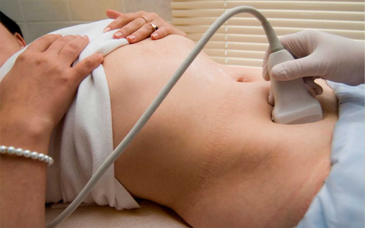
Pelvic Ultrasonography Examination: This examination can be conducted both in a transabdominal way, through the abdomen, and in a transvaginal way, through the vagina. The "doppler ultrasonographic examination" makes considerable contributions to the detailed examination of the pelvic region venous blood flow.
Pelvic computed tomography (CT) or magnetic resonance (MR) imaging: These methods are critical for the detailed demonstration and examination of pelvic region varicose veins. Both the CT and the MRI provide information on vascular structures and reveal these structures' connections with other pelvic anatomical structures such as "ovaries and uterus." Magnetic resonance imaging with contrast dye is a superior method compared to computed tomographic examination because it does not emit radiation during the analysis.
Diagnostic Venography: A venographic examination requires the imaging devices used in the angiography department. Generally, the right inguinal vein is used to perform this procedure. Left and right ovarian veins are viewed separately with the help of a wire and catheter (plastic tube) placed in the inguinal vein. In this way, the diameters of the veins and the directions of their blood flow are determined.
Laparoscopic Examination: This examination should be resorted to especially for patients with severe chronic pelvic pain. The laparoscopic analysis fails to be useful because this method is used on 80% to 90% of inpatients with pelvic varicose enlargements, and the abdomen is supplied with carbon dioxide during the application. Because this examination causes shrinkage of varicose expansions.
What are the Definitive Criteria for Diagnosing Pelvic Congestion Syndrome?
Presence of the ovarian vein larger than 4 mm in diameter
The presence of slowed blood flow (less than 3 cm/second) within the pelvic veins or the reverse venous blood flow, especially in the left ovarian vein
The presence of bilateral connections between the uterus veins (arcuate veins) and the enlarged pelvic vessels